Clinical Guide to In-Office Dermal Remodeling: Lasers, Chemical Peels & Microneedling
Topical skincare manages epidermal turnover and barrier hydration, but structural dermal remodeling requires controlled micro-injury. Understand the biophysics of acoustic, thermal, and chemical wounding, chromophore targeting, and safe execution across all Fitzpatrick phototypes.
Remodeling Physics: The Triple Wounding Principle
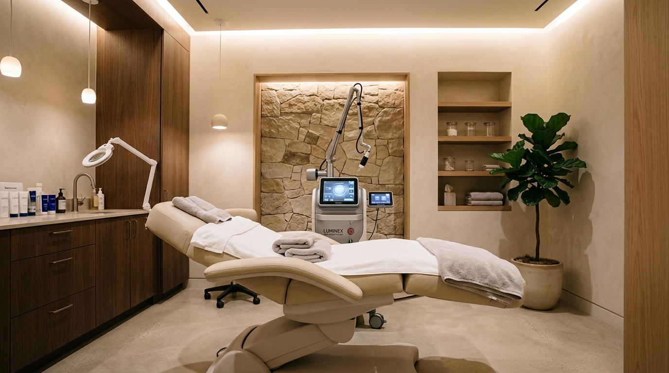
1. Dermal Remodeling Modality Spectrum & Chromophore Physics
Every in-office energy device targets a specific tissue chromophore: intracellular water, oxyhemoglobin, or melanin. Selective photothermolysis relies on matching laser wavelength to chromophore absorption while keeping pulse duration shorter than the tissue's Thermal Relaxation Time (TRT):
| Remodeling Technology | Primary Chromophore / Mechanism | Dermal Depth | Downtime Window | Fitzpatrick Safety |
|---|---|---|---|---|
| Fractional CO2 (10,600 nm) | Intracellular Water (Ablative Vaporization) | 0.5–1.5 mm | 7–14 Days | Types I–III only |
| Erbium:YAG (2,940 nm) | Water (High Absorption, Cold Ablation) | 0.2–0.8 mm | 4–7 Days | Types I–IV |
| Non-Ablative Fraxel (1550/1927 nm) | Water (Sub-surfaced MTZ Coagulation) | 0.3–1.0 mm | 2–4 Days | Types I–V |
| RF Microneedling (Morpheus8) | Ohmic Resistance (Fractional Coagulation) | 0.5–4.0 mm | 24–48 Hours | All Types (I–VI) |
| Medium-Depth TCA (20–35%) | Protein Keratocoagulation (Frosting) | 0.4–0.6 mm (Upper Reticular) | 5–8 Days | Pre-treated I–IV |
For a comprehensive comparison between radiofrequency energy and traditional acoustic stamping, consult our detailed analysis on RF Microneedling vs. Traditional Microneedling.
2. The Neocollagenesis Biology: The 180-Day Remodeling Curve
Patients frequently expect immediate structural tightening post-procedure. However, true dermal transformation follows a rigid physiological wound-healing cascade spanning six months:
Platelets release transforming growth factor-beta (TGF-β) and epidermal growth factor (EGF). Edema and erythema peak. Clinicians must avoid suppressing this acute inflammatory signaling with high-dose corticosteroids, as controlled inflammation is the necessary catalyst for collagen induction.
Dermal fibroblasts migrate into micro-coagulation zones, synthesizing glycosaminoglycans (hyaluronic acid) and fragile, pliable Type III collagen (reticulin). Re-epithelialization completes as keratinocytes bridge the epidermal basement membrane.
Lysyl oxidase enzymatically cross-links collagen fibers. Immature Type III collagen is resorbed and replaced by dense, parallel bundles of high-tensile Type I collagen. Elastin fibers are restored, manifesting as clinical lifting, pore diameter reduction, and dermal thickening.
3. The Fitzpatrick Paradox: Preventing Post-Inflammatory Hyperpigmentation
In higher Fitzpatrick phototypes (Types IV to VI—common in Mediterranean, Hispanic, South Asian, and African ancestries), melanocytes in the basal epidermal layer are hyper-reactive. When thermal laser heat diffuses into the dermo-epidermal junction, it triggers melanocyte-stimulating hormone (MSH) and endothelin-1, inducing stubborn Post-Inflammatory Hyperpigmentation (PIH).
To prevent PIH disasters:
- Pre-Conditioning Tyrosinase Inhibitors: For Fitzpatrick IV–VI, prime the skin for 4 to 6 weeks pre-procedure using Hydroquinone (4%), Tranexamic Acid (3–5%), Azelaic Acid (15%), and Kojic Acid to quiesce melanogenesis.
- Insulated Needle Architecture: In RF microneedling, always utilize insulated needles where the needle shaft is coated in biocompatible silicone, restricting RF thermal emission strictly to the distal 0.3mm needle tip situated safely within the reticular dermis, bypassing the melanin-rich epidermis completely.
- Peel Depth Control: Understand the chemical mechanics of peeling agents; explore our clinical protocol on Medium-Depth TCA Peels & PIH Management.
4. Multi-Modality Stacking: Energy Devices, Peels & Neuromodulators
Elite aesthetic clinics execute synergized treatment roadmaps:
- Same-Day Stacking: RF microneedling combined with non-crosslinked hyaluronic acid and exogenous platelet-rich plasma (PRP) immediately post-wounding maximizes active transport through open micro-conduits.
- Neuromodulator Timing: Always administer botulinum toxin (Botox, Dysport, Daxxify) either 2 weeks prior to energy treatments or 48 hours post-procedure. Injecting neuromodulators immediately prior to heat-generating lasers causes thermal spread and migration of the toxin into adjacent unintended musculature. Learn how to extend neurotoxin duration in Neuromodulator Longevity & Zinc Bioavailability.
Frequently Asked Questions: Dermal Remodeling Procedures
What is the difference between ablative and non-ablative laser resurfacing?
Ablative lasers (CO2 10,600nm, Er:YAG 2,940nm) vaporize the external epidermal layer while heating the underlying dermis, yielding profound collagen restructuring at the cost of 7 to 14 days of exudative downtime and higher post-inflammatory hyperpigmentation risk. Non-ablative fractional lasers (1,550nm Erbium-glass, 1,927nm Thulium) heat dermal columns while leaving the stratum corneum intact, requiring 3 to 5 sessions with only 24 to 72 hours of mild erythema.
Which med-spa treatments are safe for Fitzpatrick skin types IV to VI?
For melanated skin (Fitzpatrick IV to VI), insulated radiofrequency microneedling (such as Morpheus8 with silicone-coated needles), 1064nm Nd:YAG lasers, and superficial mandelic or low-concentration lactic acid peels are safest. These modalities bypass epidermal melanin absorption, delivering energy directly to the reticular dermis without triggering post-inflammatory hyperpigmentation (PIH).
How many weeks should you wait between chemical peels and microneedling sessions?
Clinicians recommend waiting a minimum of 4 to 6 weeks between deep dermal remodeling sessions. This matches the biological neocollagenesis timeline, where fibroblast proliferation peaks at day 21 and the transition from immature Type III collagen to tensile Type I collagen requires 6 to 12 weeks of uninhibited cellular remodeling.