Epidermal Growth Factors (EGF) & SH-Oligopeptide-1: Molecular Kinetics & Cellular Safety
Since Dr. Stanley Cohen's Nobel Prize-winning discovery of Epidermal Growth Factor in 1986, biotechnology has sought to harness recombinant polypeptides for cellular regeneration. Marketed under the INCI designation sh-Oligopeptide-1, synthetic human EGF represents one of the most potent mitogenic agents in modern cosmeceuticals. Yet its clinical utilization is fraught with formulation dilemmas: how does a 6.2-kilodalton macromolecule traverse the stratum corneum, and does chronic topical receptor stimulation raise oncogenic risks?
1. Structural Biology: Deconstructing sh-Oligopeptide-1 vs. Cosmetic Peptides
In cosmetic labeling, the prefix "sh-" designates synthetic human. Recombinant human EGF (rhEGF) is synthesized by inserting the exact cDNA sequence of human epidermal growth factor into microbial expression vectors—predominantly Escherichia coli, bioengineered yeast (Pichia pastoris), or microalgae. Following fermentation, the protein is harvested and chromatographically purified to yield an identical chemical match to endogenous human EGF.
Chemically, sh-Oligopeptide-1 is a compact, single-chain globular polypeptide comprising 53 amino acid residues with a molecular weight of approximately 6,215 Daltons (~6.2 kDa):
H-Asn-Ser-Asp-Ser-Glu-Cys-Pro-Leu-Ser-His-Asp-Gly-Tyr-Cys-Leu-His-Asp-Gly-Val-Cys-Met-Tyr-Ile-Glu-Ala-Leu-Asp-Lys-Tyr-Ala-Cys-Asn-Cys-Val-Val-Gly-Tyr-Ile-Gly-Glu-Arg-Cys-Gln-Tyr-Arg-Asp-Leu-Lys-Trp-Trp-Glu-Leu-Arg-OH
Formulators frequently confuse true growth factors with generic cosmetic peptides. Synthetic oligopeptides (such as Palmitoyl Tripeptide-1 or Acetyl Hexapeptide-8) are tiny oligomers of 3 to 8 amino acids attached to lipophilic fatty acid tails to enhance passive delivery. They act as weak biomimetic analogues. In stark contrast, sh-Oligopeptide-1 is a full-sized biologically active protein capable of triggering irreversible intracellular kinase cascades.
2. Receptor Kinetics: The EGFR/ErbB-1 Signaling Machinery
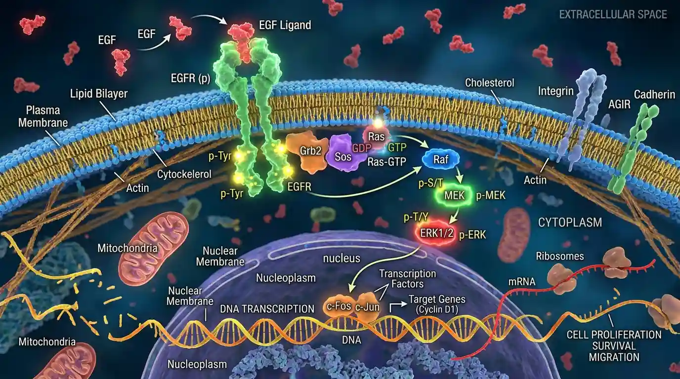
EGF exerts its biological function by binding with picomolar affinity to the extracellular domain of the Epidermal Growth Factor Receptor (EGFR / ErbB-1 / HER1). EGFR is a 170-kDa transmembrane glycoprotein abundant on basal keratinocytes, hair follicle outer root sheaths, and dermal fibroblasts.
Upon ligand engagement, the following intracellular cascade takes place within seconds:
- Receptor Dimerization: EGF binding induces conformational unmasking of the receptor dimerization arm, prompting EGFR to pair with another EGFR monomer (homodimer) or an ErbB-2/HER2 co-receptor (heterodimer).
- Tyrosine Kinase Trans-Phosphorylation: Dimerization aligns the intracellular kinase domains, causing reciprocal phosphorylation of key tyrosine residues (Tyr992, Tyr1045, Tyr1068, Tyr1173) in the cytoplasmic tail.
- The MAPK / ERK Mitogenic Cascade: Phospho-tyrosines serve as high-affinity docking sites for the adapter protein Grb2 and the guanine nucleotide exchange factor Sos. Sos converts membrane-bound Ras-GDP into active Ras-GTP, initiating the sequential kinase relay:
Ras → Raf → MEK1/2 → ERK1/2. Phosphorylated ERK translocates into the nucleus to activate immediate-early transcription factors c-Fos and c-Jun (AP-1 complex), upregulating Cyclin D1 and driving quiescent G0 keratinocytes into active S-phase DNA synthesis. - The PI3K / Akt Survival Pathway: Simultaneously, EGFR activates Phosphoinositide 3-Kinase (PI3K), which phosphorylates PIP2 into PIP3, recruiting Akt (Protein Kinase B). Activated Akt represses pro-apoptotic proteins (Bad, Bax, Caspase-9), conferring cellular resilience against oxidative stress and photo-damage.
In acute dermal injury, this synchronized biochemical storm rapidly accelerates re-epithelialization, mobilizes keratinocyte edge migration, and stimulates secondary release of basic Fibroblast Growth Factor (bFGF) and Transforming Growth Factor-beta (TGF-β) to initiate provisional matrix deposition.
3. The Penetration Paradox: How Does a 6.2 kDa Molecule Navigate the Stratum Corneum?
In transdermal pharmacology, the foundational barrier law is Bos and Meinardi’s 500-Dalton Rule. Intact human stratum corneum presents a tightly packed, hydrophobic lipid bilayer consisting of ceramides, cholesterol, and free fatty acids that virtually bars the passive diffusion of polar hydrophilic molecules exceeding 500 Da.
With a molecular mass of 6,215 Da—over twelve times the theoretical cutoff—how can topical EGF produce meaningful anti-aging or regenerative benefits in intact human skin?
On Intact Epidermis:
Passive transcellular and intercellular diffusion of intact 6.2 kDa EGF is negligible. Penetration is restricted to:
- Follicular Shunt Transport: Microscopic entry through hair follicle infundibula and sebaceous duct openings where the cornified barrier is thinned.
- Superficial Paracrine Signaling: Interaction with trace surface receptors on the outermost living keratinocytes in micro-fissured or dehydrated barriers.
- Vehicle Effect: Commercial serums often rely heavily on the occlusive and humectant properties of hyaluronic acid vehicles rather than direct protein diffusion.
On Compromised / Micro-Channeled Skin:
When the physical stratum corneum barrier is mechanically or thermally disrupted, macromolecular restrictions vanish:
- Microneedling Pores: Medical-grade microneedling (0.5 mm to 1.5 mm) punches transient micro-channels directly into the viable epidermis and papillary dermis.
- Fractional Laser Ablation: Microscopic treatment zones (MTZs) allow liquid suspensions of rhEGF to flood the wound bed.
- Receptor Saturation: Direct contact with basal keratinocyte and dermal fibroblast EGFRs delivers massive, clinically validated acceleration of re-epithelialization.
Consequently, formulating chemists agree: applying rhEGF as a conventional daily anti-aging cream yields modest results compared to its profound efficacy when deployed immediately post-procedure. To understand how molecular mass dictates topical active sequencing, review our foundational pillar, The Molecular Weight Hierarchy of Topical Actives.
4. The Oncogenesis Controversy: Does Topical EGF Promote Malignancy?
The most contentious debate surrounding sh-Oligopeptide-1 centers on cellular proliferation safety. In clinical oncology, the Epidermal Growth Factor Receptor is infamous: EGFR gene amplification and receptor overexpression are hallmark drivers in cutaneous squamous cell carcinoma (cSCC), basal cell carcinoma (BCC), glioblastoma, and metastatic lung adenocarcinoma. Targeted cancer therapies (e.g., cetuximab, erlotinib) are designed specifically to inhibit EGFR activity.
This biological reality raises an essential clinical question: could chronic daily application of an EGFR ligand inadvertently stimulate dormant pre-malignant clones or accelerate subclinical keratinocyte carcinomas?
What the Clinical Evidence Demonstrates
Multiple robust toxicology evaluations and clinical trials offer clear reassurance regarding healthy skin:
- Non-Mutagenic Profile: Recombinant human EGF is a mitogen, not a mutagen. It does not damage DNA, cause chromosomal aberrations, or transform healthy cells into cancerous genotypes. It only accelerates the mitotic cycle of cells already expressing normal receptor physiology.
- The Radiation Dermatitis Cohort: In a landmark multicenter study involving 1,172 oncology patients undergoing radiation therapy for malignant solid tumors, topical rhEGF cream was administered daily to irradiated fields to treat acute radiation dermatitis. Across the entire cohort, EGF accelerated wound closure without increasing tumor recurrence, local progression, or secondary skin cancer incidence.
- Negative In-Vitro Carcinogenesis Screens: Standard cosmeceutical testing on human epidermal models confirms that cosmetic concentrations of sh-Oligopeptide-1 (typically between 0.0001% and 0.001%, or 1 to 10 ppm) fail to elicit invasive migratory phenotypes in non-tumorigenic keratinocytes.
Prudent Clinical Restrictions: The Atelier Safety Rule
While topical EGF has never been proven to generate de novo skin tumors, its mitogenic potency requires conservative clinical boundaries. Aesthetic specialists advise strict precautionary avoidance under the following circumstances:
- × Never apply over diagnosed skin cancers: Avoid application over active Basal Cell Carcinomas, Squamous Cell Carcinomas, or Melanomas.
- × Precancerous Dysplasia: Individuals with multiple Actinic Keratoses (AKs) or a personal history of field cancerization should avoid chronic daily EGF usage.
- × Atypical Nevi: Avoid direct, repeated application over dysplastic or rapidly changing melanocytic nevi.
5. Formulation Chemistry: Stability, Denaturation & Layering Rules
Because sh-Oligopeptide-1 is an authentic protein rather than a simple acid or synthetic humectant, its biological activity is exceptionally vulnerable to environmental degradation. In the formulation laboratory, preserving rhEGF stability demands strict adherence to three chemical principles:
- The pH Denaturation Threshold: EGF maintains its tertiary disulfide-bonded conformation strictly between pH 5.5 and 7.5. Exposing EGF to acidic serums—such as unbuffered 15% L-Ascorbic Acid (pH 2.8–3.2) or 7% Glycolic Acid (pH 3.5)—protonates aspartic and glutamic acid side chains, unfolding the protein within seconds. Always apply direct acids first, allow a complete neutralization window, or reserve EGF exclusively for alternate evenings.
- Thermal & Proteolytic Sensitivity: Unlike classic retinoids, which degrade primarily via photo-oxidation, EGF polypeptides are digested by endogenous cutaneous proteases and denatured by excessive heat. Formulations must incorporate stabilizing polyols (trehalose, mannitol) and cold-chain or vacuum-sealed airless dispensing.
- Peptide Layering Hierarchy: EGF pairs exquisitely with non-acidic signal messengers such as copper tripeptides and multi-molecular hyaluronic acid. However, direct metal ions in unchelated formulas can interfere with disulfide bridges. Learn more about metal chelation risks in our guide on Copper Peptides vs. Direct Acids: Denaturing & Incompatibility.
When integrating growth factors with cellular renewal agents, formulators recommend alternating nights with gentle retinoid cascades; review the full biochemical pathway in The Retinoid Conversion Cascade: Retinal, Retinol & HPR. For medical-grade collagen induction protocols, explore our comparative analysis on RF Microneedling vs. Traditional Microneedling.
Frequently Asked Questions: EGF & SH-Oligopeptide-1
Can a 6,200 Dalton protein like EGF penetrate intact human skin?
Passive transdermal penetration of intact stratum corneum is strictly governed by the 500-Dalton rule. At approximately 6,200 Daltons (6.2 kDa), full-length recombinant EGF cannot diffuse freely through the compact intercellular lipid bilayer. On uncompromised skin, biological action is limited to superficial keratinocyte signaling and follicular shunt pathways. However, following clinical microneedling or fractional laser passes, micro-channels bypass the stratum corneum, enabling direct EGFR binding in the viable epidermis and dermis.
Does topical Epidermal Growth Factor cause or accelerate skin cancer?
Extensive clinical reviews and large multicenter oncology trials (including cohort studies in radiation dermatitis patients) demonstrate no evidence that topical recombinant human EGF initiates oncogenesis or induces de novo malignant transformations in healthy human skin. However, because EGF is a potent mitogen that activates EGFR-dependent cell division, it is clinically contraindicated over active malignancies, precancerous actinic keratoses, or recent melanoma excision sites.
What is the biochemical difference between sh-Oligopeptide-1 and cosmetic peptides like Matrixyl?
Cosmetic peptides like palmitoyl pentapeptide-4 (Matrixyl) are short synthetic fragments consisting of 3 to 6 amino acids that act as subtle biomimetic messengers. In contrast, sh-Oligopeptide-1 is a complete, full-length recombinant human protein of 53 amino acids with three native disulfide bonds that binds specifically to transmembrane EGFR (ErbB-1) receptors to initiate MAPK/ERK and PI3K/Akt kinase phosphorylation cascades.
Can you use Epidermal Growth Factors in the same routine as direct acids or retinoids?
Strong direct chemical exfoliants (glycolic or salicylic acid below pH 4.0) denature the tertiary three-dimensional protein fold of EGF, hydrolyzing its disulfide bonds and rendering it biologically inert. While retinoids can be cycled with EGF on alternating evenings, growth factors should never be mixed in the same application phase with low-pH unbuffered acids.