IPL Photofacial vs. Pulsed Dye Laser: Chromophore Physics of Erythema & Melanin
Facial photodamage rarely presents in isolation. Chronic ultraviolet exposure generates a complex clinical tapestry: dilated superficial microvasculature, persistent erythematotelangiectatic rosacea, mottled epidermal solar lentigines, and poikiloderma of Civatte. In aesthetic laser physics, clinical efficacy depends upon selective photothermolysis—matching precise optical wavelengths and pulse durations to specific cutaneous chromophores. Unpack the biophysical differences between broadband Intense Pulsed Light (IPL) and the 595 nm monochromatic Pulsed Dye Laser (PDL).
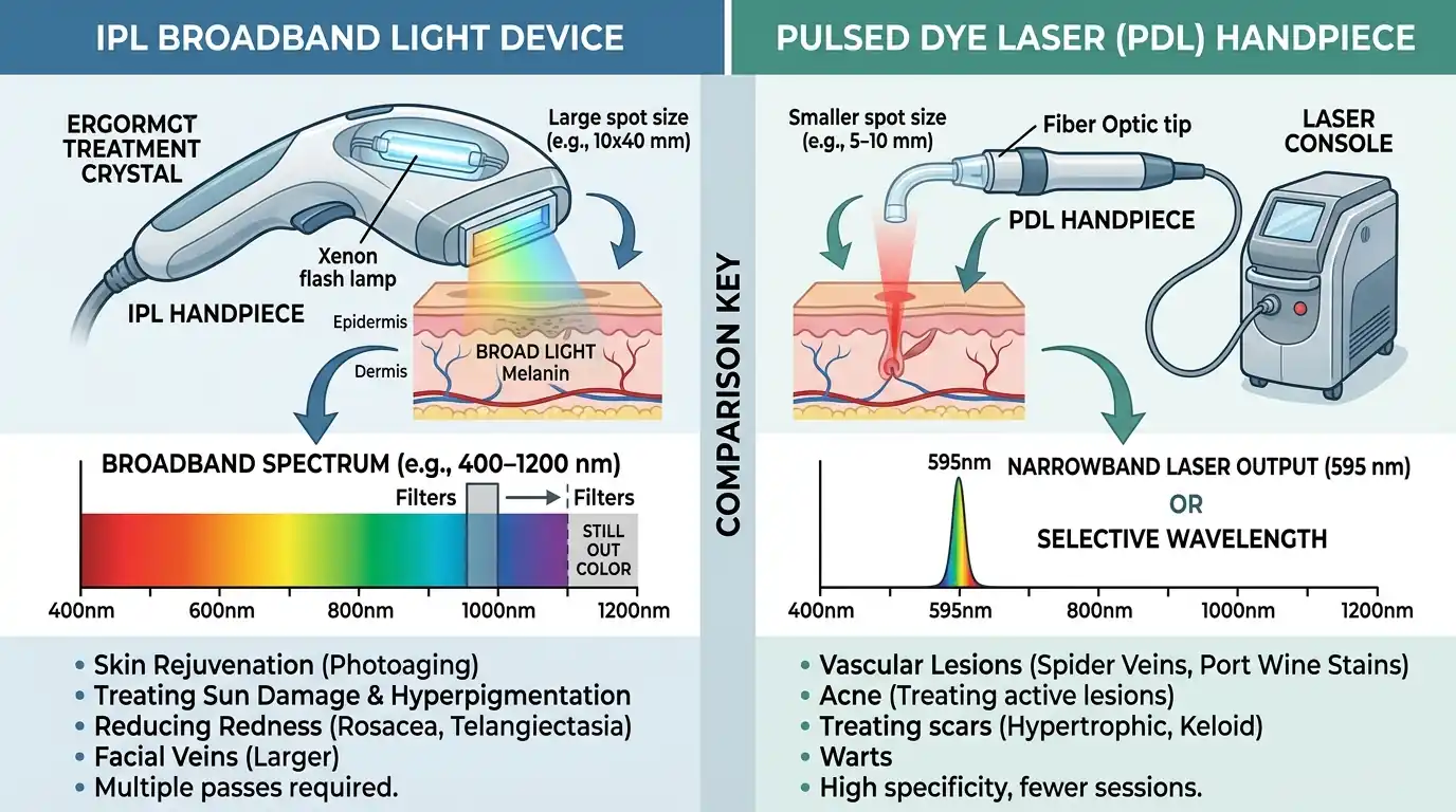
1. Fundamental Physics: Polychromatic Flashlamp vs. Monochromatic Laser
The most fundamental misconception among aesthetic patients is classifying Intense Pulsed Light as a "laser." In optical physics, true lasers and IPL devices occupy entirely different technological paradigms:
Intense Pulsed Light (IPL)
- Optical Nature: Polychromatic, non-coherent, divergent broadband radiation emitted by a high-output Xenon flashlamp.
- Emission Spectrum: 500 nm to 1,200 nm, sculpted by interchangeable optical cutoff filters (e.g., 515 nm, 560 nm, 590 nm) that block shorter, burning wavelengths.
- Spot Architecture: Large sapphire or quartz coupling blocks (typically 10 × 30 mm to 15 × 35 mm), enabling rapid treatment of full facial, chest, and dorsal hand surfaces.
Pulsed Dye Laser (PDL)
- Optical Nature: Monochromatic, spatially and temporally coherent, collimated beam produced by rhodamine organic dye excited by a flashlamp.
- Single Wavelength: Exactly 595 nm (the modern Candela Vbeam standard, superseding older 585 nm systems).
- Spot Architecture: Circular collimated spots (3 mm to 12 mm) with flat-top or Gaussian energy profiles, engineered for surgical microvascular tracing.
Because IPL emits multiple wavelengths simultaneously, photon energy distributes across multiple competing chromophores in the skin—primarily melanin, hemoglobin, and to a lesser extent water. In contrast, PDL channels its entire photon energy into a single razor-sharp optical peak.
2. Chromophore Absorption Dynamics: The Hemoglobin-Melanin Competition
To destroy abnormal blood vessels or hyperpigmented macules without causing non-specific thermal scarring of surrounding collagen, Anderson and Parrish’s principle of selective photothermolysis dictates that the target chromophore must absorb light far more intensely than surrounding skin constituents.
In cutaneous optics, two primary chromophores compete for visible light:
Conversely, the absorption spectrum of melanin drops continuously and monotonically from ultraviolet through infrared. In light Caucasian skin (Fitzpatrick I–II), low epidermal melanin allows 500–600 nm light to reach vascular targets unimpeded. However, in darker skin types (Fitzpatrick IV–VI), epidermal melanin acts as an aggressive competing optical shield, absorbing yellow light and converting it into dangerous heat at the dermal-epidermal junction.
3. Thermal Relaxation Time (TRT): Purpuric vs. Sub-Purpuric Vascular Coagulation
The Thermal Relaxation Time (TRT) represents the duration required for a heated chromophore to lose 50% of its thermal energy to surrounding tissue via conduction. TRT is proportional to the square of the target structure's diameter:
- Microscopic Melanosomes (0.5–1 μm): TRT of approximately 50 to 250 nanoseconds (best cleared by Q-switched or picosecond lasers, or millisecond IPL with gentle micro-crusting).
- Capillary Ectasias & Telangiectasias (10–100 μm): TRT between 1 and 10 milliseconds.
- Venules & Deep Port-Wine Stain Vessels (100–300 μm): TRT between 10 and 50 milliseconds.
The Purpuric Threshold in PDL Physics
Historically, pulsed dye lasers operated with ultra-short pulse durations (0.45 to 1.5 ms). When high fluences are delivered faster than the vessel’s TRT, intravascular blood undergoes explosive boiling and rapid photomechanical vaporization. The vessel wall ruptures violently, causing extensive extravasation of erythrocytes into the dermis. This manifests clinically as dark, round, purple bruises—purpura—that persist for 10 to 14 days. While purpuric settings remain unmatched for clearing stubborn pediatric port-wine stains, modern cosmetic adult patients routinely refuse weeks of visible facial bruising.
Modern 595 nm PDL devices overcome this limitation through extended pulse durations (6 ms, 10 ms, 20 ms, up to 40 ms) coupled with millisecond-timed cryogen spray cooling (Dynamic Cooling Device / DCD). By delivering energy over a prolonged window, the laser gently coagulates intravascular oxyhemoglobin into methemoglobin and induces slow vessel wall collapse without rupture. This sub-purpuric protocol yields dramatic rosacea erythema reduction with zero bruising and less than 48 hours of mild swelling.
4. Clinical Head-to-Head: Matching the Technology to the Pathology
Selecting between an IPL photofacial and a pulsed dye laser requires an exacting diagnostic evaluation of the patient’s primary cosmetic complaint:
| Clinical Indication | IPL Photofacial (500–1200 nm) | Pulsed Dye Laser (595 nm) |
|---|---|---|
| Discrete Nasal & Cheek Telangiectasias | Moderate clearance; requires small tracing filters | Gold Standard; instantaneous vessel closure |
| Diffuse Erythematotelangiectatic Rosacea | Excellent; covers full face rapidly with large spot | Excellent; superior depth of vessel coagulation |
| Solar Lentigines ("Sun Spots / Age Spots") | Superior; darkens and sloughs pigment via "coffee-grounds" | Ineffective; 595 nm bypasses epidermal melanin clusters |
| Poikiloderma of Civatte (Lateral Neck) | Premier Choice; treats red vessels and brown mottled pigment simultaneously | Clears red component only; leaves brown pigment untouched |
| Port-Wine Stains & Hemangiomas | Sub-optimal; insufficient coherent energy density | Undisputed Medical Gold Standard |
| Typical Number of Sessions | 3 to 5 sessions spaced 4 weeks apart | 2 to 4 sessions spaced 4 to 6 weeks apart |
For patients presenting with classic photoaging—a mottled mixture of broken capillaries, diffuse cheek flushing, and discrete brown lentigines across the forehead and temples—broadband IPL represents the single most cost-effective and comprehensive modality. Conversely, for the patient with pure vascular rosacea, inflammatory acne erythema, or post-surgical scars, the 595 nm PDL provides surgical precision without risking excessive epidermal melanin absorption.
5. Safety Protocols in Fitzpatrick Types IV–VI: Preventing Thermal Blistering & PIH
Performing optical light procedures on darker skin phototypes requires exceptional clinical vigilance. Because epidermal basal layers in Fitzpatrick IV–VI skin contain high densities of eumelanin, applying short-wavelength light (515–560 nm) creates catastrophic surface heat:
- The Risk of Striping and Footprints: Non-uniform IPL contact or inadequate coupling gel results in square, hyperpigmented burn marks ("footprinting") that take 6 to 12 months to fade.
- Long-Wavelength Cutoff Filters: When treating skin type IV with IPL, operators must shift cutoff filters to 590 nm, 615 nm, or 640 nm. This eliminates the short green wavelengths that burn melanin, allowing yellow and near-infrared light to safely penetrate deeper into the dermis.
- Cryogen Pre-Cooling with PDL: Modern 595 nm PDL systems utilize millisecond bursts of cryogen spray (1,1,1,2-tetrafluoroethane) delivered 20 to 40 milliseconds before the laser pulse. This flash-freezes the superficial epidermis to approximately 4°C, insulating melanin from thermal injury while the laser pulse passes through to coagulate underlying blood vessels.
Prior to undergoing either treatment, patients prone to pigmentary rebound should prime their skin for 4 weeks with tyrosinase-inhibiting cosmeceuticals; learn more in our detailed protocol on TCA Peels & Melasma PIH Management. For non-optical collagen induction that completely avoids melanin competition, consider non-laser modalities outlined in RF Microneedling vs. Traditional Microneedling and our comprehensive overview in Clinical In-Office Skin Remodeling.
Frequently Asked Questions: IPL vs. Pulsed Dye Laser
Is Pulsed Dye Laser or IPL more effective for facial rosacea and telangiectasias?
Pulsed Dye Laser (595 nm) is the clinical gold standard for pure vascular pathologies, including stubborn telangiectasias, cherry angiomas, and port-wine stains. Because it emits a coherent, monochromatic 595 nm beam precisely aligned with oxyhemoglobin's beta absorption plateau, it destroys blood vessels with minimal scatter. However, for generalized photoaging involving both diffuse facial redness and brown epidermal sun spots (solar lentigines), broadband IPL is frequently preferred for its dual-chromophore clearance in a single session.
What is the difference between purpuric and sub-purpuric PDL settings?
Purpuric PDL settings utilize short pulse durations (0.45 to 1.5 milliseconds) that heat vessels faster than their thermal relaxation time, causing instantaneous microvascular mechanical rupture, extravasation of red blood cells, and dark purple bruising lasting 7 to 14 days. Modern sub-purpuric settings utilize extended pulse durations (6 to 40 milliseconds) with dynamic cryogen spray cooling to slowly coagulate vessel walls without rupture, delivering vascular clearance with zero bruising and minimal social downtime.
Can IPL or PDL be safely performed on Fitzpatrick Skin Types IV to VI?
Darker phototypes (Fitzpatrick IV–VI) possess high concentrations of epidermal melanin that compete with hemoglobin for photon absorption between 500 nm and 600 nm. Broadband IPL carries substantial risk of epidermal burns, blistering, and post-inflammatory hyperpigmentation (PIH) unless long-wavelength cutoff filters (≥590–640 nm) and conservative fluences are strictly applied. Sub-purpuric 595 nm PDL equipped with millisecond cryogen cooling is generally safer for vascular targets in Fitzpatrick IV, but treatments in Fitzpatrick V and VI require extreme caution or alternative Nd:YAG 1064 nm wavelengths.
How does the post-treatment healing process differ between IPL and PDL?
Following an IPL photofacial, targeted epidermal melanin darkens within 24 hours into micro-crusts resembling "coffee grounds" or dark pepper specks, which flake off naturally over 5 to 7 days without open wounds. Following sub-purpuric PDL, patients experience transient facial edema and mild erythema resolving within 24 to 48 hours; if purpuric settings are selected, distinct purple-black circular bruises persist for 7 to 10 days.